Fontanels
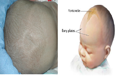
Fontanelles are soft spots on a baby's head which, during birth, enable the bony plates of the skull to flex, allowing the child's head to pass through the birth canal. The ossification of the bones of the skull causes the fontanelles to close over by 18 to 24 months. The closures eventually form the sutures of the neurocranium. Other than the anterior and posterior fontanelles, the mastoid fontanelle and the sphenoidal fontanelle are also significant.
Anterior fontanelleThe skull of a newborn consists of five main bones: two frontal bones, two parietal bones, and one occipital bone. These are joined by fibroussutures, which allow movement that facilitates childbirth and brain growth.
Anterior fontanelleThe skull of a newborn consists of five main bones: two frontal bones, two parietal bones, and one occipital bone. These are joined by fibroussutures, which allow movement that facilitates childbirth and brain growth.
- At birth, the skull features a small posterior fontanelle, an open area covered by a tough membrane, where the two parietal bones adjoin the occipital bone (at the lambda). This fontanelle usually closes during the first two to three months of an infant's life. This is called intramembranous ossification. The mesenchymal connective tissue turns into bone tissue.
- The much larger, diamond-shaped anterior fontanelle where the two frontal and two parietal bones join generally remains open until the child is about two years of age, however, in cleidocranial dysostosis it is often late in closing or may never close. The anterior fontanelle is useful clinically. Examination of an infant includes palpating the anterior fontanelle.
- Two smaller fontanelles are located on each side of the head, more anteriorly the sphenoidal (between the sphenoid, parietal, temporal, and frontal bones) and more posteriorly the mastoid (between the temporal, occipital, and parietal bones).
